
Do you know how many ultrasounds during pregnancy are required?
For most expectant parents, few moments in a pregnancy journey are as moving as seeing their baby on screen for the very first time. Ultrasound scans have transformed prenatal care, giving parents and healthcare teams a real-time view inside the womb. Parents often wonder about the right number of scans and when to expect each one — and the answer depends on several factors, including the mother’s overall health, her ob-gyn’s recommendations, and whether her pregnancy is considered low-risk or high-risk.
What Are Ultrasound Scans and How Do Sound Waves Work?
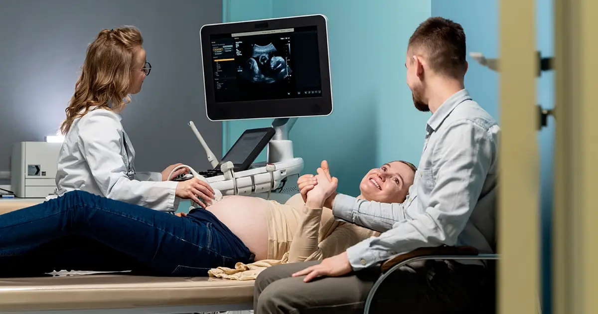
Ultrasound scans use high-frequency sound waves to produce real-time images of the baby developing inside the uterus. As the sound waves bounce off tissues and organs, they return as signals that are converted into images on a screen.
Two main types of ultrasound are used in standard prenatal care:
- Abdominal ultrasound: A transducer is moved across the abdomen using gel. A full bladder may be needed early in pregnancy for clearer imaging.
- Transvaginal ultrasound: A small probe is gently inserted into the vagina. This is especially useful in the first trimester when the uterus is still small.
Both help the ob-gyn and healthcare provider monitor the baby’s growth, assess potential problems, and support the developing baby throughout the pregnancy.
How Many Ultrasounds Is Standard? The Ob-Gyn and Healthcare Provider’s Role
The American College of Obstetricians and the American Institute of Ultrasound in Medicine are widely referenced for pregnancy imaging guidance. For most pregnant women carrying a healthy pregnancy without complications, two standard ultrasounds — one early scan and one mid-pregnancy anatomy scan — form the baseline.
The number of ultrasounds a woman receives is shaped by her individual risk factors and medical history. For most, two to three ultrasound scans throughout the full term are considered routine. The ob-gyn and healthcare provider together determine what is most appropriate for each person.
First Trimester Ultrasound: The Dating Scan and Early Ultrasound
The first ultrasound is typically scheduled between six and ten weeks of pregnancy. This early ultrasound is commonly called the dating scan or dating ultrasound, as it helps confirm the due date and verifies that the pregnancy is developing normally.
The first trimester scan confirms the gestational sac, verifies the fetal heartbeat, and rules out an ectopic pregnancy — a serious condition where the embryo implants outside the uterus.
In some cases, the healthcare provider may recommend blood tests or additional screening procedures such as chorionic villus sampling.
Parents can use the Pregnancy Due Date Calculator at MyWellCal to easily estimate their due date based on their weeks of pregnancy.
Second Trimester: Baby’s Development and the Anatomy Scan
Around 18 to 20 weeks, most ob-gyns schedule the mid-pregnancy anatomy scan. This is one of the most detailed ultrasound examinations in the pregnancy journey, and it covers a wide range of information about the baby.
The anatomy scan examines baby’s organs, baby’s size, and the location of the placenta. It screens for birth defects and can reveal the baby’s sex if parents wish to know.
During this ultrasound exam, the ob-gyn and healthcare provider will also review:
- Baby’s growth and baby’s position within the uterus
- Position of the placenta (to identify placenta previa, where the placenta blocks the cervix)
- Amount of amniotic fluid surrounding the baby
- Blood flow through the umbilical cord via Doppler ultrasound
- Baby’s heartbeat and heart rate
This scan often produces the clearest images of your baby that parents will treasure for years to come.
Third Trimester Ultrasound: Tracking Fetal Growth and Well-Being
A Third Trimester Ultrasound is usually scheduled between 32 and 36 weeks. At this stage of pregnancy, the healthcare team focuses on fetal growth, baby’s position, and amniotic fluid levels.
If there are concerns about the growth rate or health of the placenta, additional ultrasounds may be ordered in the third trimester. Monitoring the well-being of both mother and baby remains the central goal at this stage.
For women managing health conditions, the third trimester may involve more frequent pregnancy scans to ensure everything is progressing as expected.

High-Risk Pregnancies and the Need for Additional Ultrasounds
Women with high-risk pregnancies typically undergo significantly more ultrasound scans than those in low-risk situations. A pregnancy may be classified as high-risk due to:
- High blood pressure or preeclampsia
- Gestational diabetes
- Medical conditions such as thyroid disorders or heart disease
- Concerns about fetal growth or the development of the fetus
- A medical reason linked to previous birth defects or pregnancy complications
- Multiple pregnancies (twins, triplets, or more)
In high-risk pregnancies, healthcare professionals may recommend ultrasound examinations every two to four weeks. Diagnostic tests such as 3D ultrasound and Doppler ultrasound are more commonly used to assess specific risk factors and the baby’s health.
Abdominal Ultrasound and Transabdominal Ultrasounds: Types of Ultrasound Explained
Several different technologies are used during pregnancy depending on the week ultrasound findings and the clinical need. The type of ultrasound selected varies based on what the medical team needs to assess:
- Standard abdominal imaging: The most commonly performed procedure across all trimesters.
- Transvaginal ultrasound: Preferred for early ultrasound imaging when detailed close-up views are needed.
- Doppler ultrasound: Monitors blood circulation and baby’s health.
- 3D ultrasound: Provides detailed views of the baby’s development and growth and development of key structures.
Each type of ultrasound serves a distinct clinical purpose and is chosen based on the specific needs of the pregnancy.
What to Expect During an Ultrasound Exam
During a standard ultrasound exam, an ultrasound technician applies warm gel to the abdomen and gently moves the transducer across the skin. The technology produces real-time images on the screen within minutes.
The process is entirely painless and typically takes between 20 and 45 minutes.
Ultrasound results are reviewed by the medical team and discussed with parents. If any concern is identified, a follow-up pregnancy scan or referral to a health professional may be recommended.
The Role of Healthcare, Prenatal Care, and Women’s Health
Regular check-ups with the healthcare team form the backbone of a healthy pregnancy. From the pregnancy test that first confirms new life to the final prenatal care appointment before delivery, every step matters.
Ultrasound examinations are just one part of a broader healthcare approach that includes blood tests, physical exams, and close monitoring of the expectant mother’s overall health.
A committed women’s health team can guide parents through every trimester with confidence, ensuring that each scan contributes to a complete picture of how the pregnancy is progressing.
From First Ultrasound to Final Pregnancy Scan: What Every Parent Should Know
Understanding how ultrasound scans reveal information at each stage gives expectant parents both clarity and peace of mind. Each scan builds a fuller picture of the health of your baby and ensures any concerns are caught early.
For women with high-risk pregnancies, more frequent imaging through ultrasound scans provides important reassurance and allows healthcare professionals to act quickly when needed.For those tracking key milestones and estimating important dates, the MyWellCal Pregnancy Due Date Calculator is a great starting point. For more resources on women’s health and pregnancy guidance, visit MyWellCal.
External Resources
For medically reviewed information, readers can visit ACOG – Ultrasound Exams, AIUM – Obstetric Ultrasound, and Mayo Clinic – Fetal Ultrasound
Frequently Asked Questions
How many ultrasounds are normal during pregnancy?
When is the first ultrasound done?
Do all pregnant women get a third trimester ultrasound?
Why would I need extra ultrasounds during pregnancy?
Is ultrasound safe during pregnancy?
Our Fact Checking Process
We prioritize accuracy and integrity in our content. Here's how we maintain high standards:- Expert Review: All articles are reviewed by subject matter experts.
- Source Validation: Information is backed by credible, up-to-date sources.
- Transparency: We clearly cite references and disclose potential conflicts.
Our Review Board
Our content is carefully reviewed by experienced professionals to ensure accuracy and relevance.- Qualified Experts: Each article is assessed by specialists with field-specific knowledge.
- Up-to-date Insights: We incorporate the latest research, trends, and standards.
- Commitment to Quality: Reviewers ensure clarity, correctness, and completeness.